May 31, 2013:
|
If you’re interested in microbiology, you are probably already reading Elio Schaechter’s blog Small Things Considered. It is hosted by the American Society for Microbiology. If you are not reading it yet, here is a great reason to start: Art on a Dish.
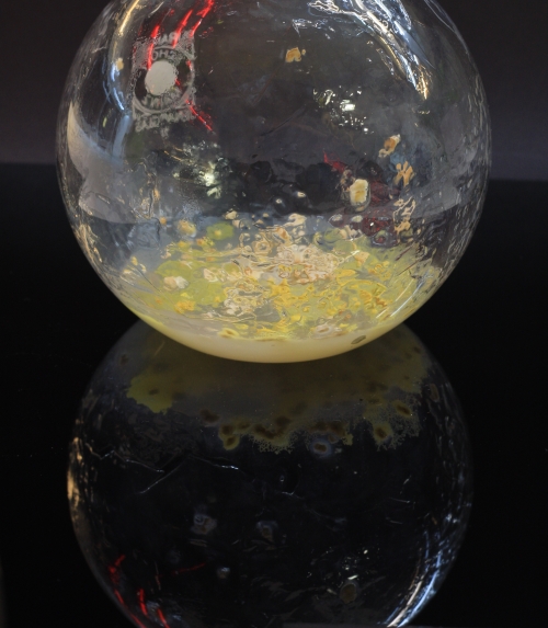
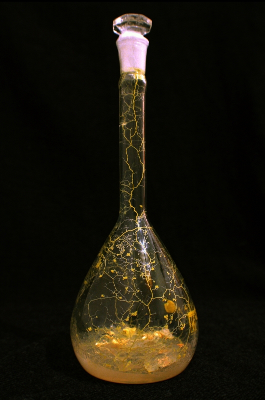
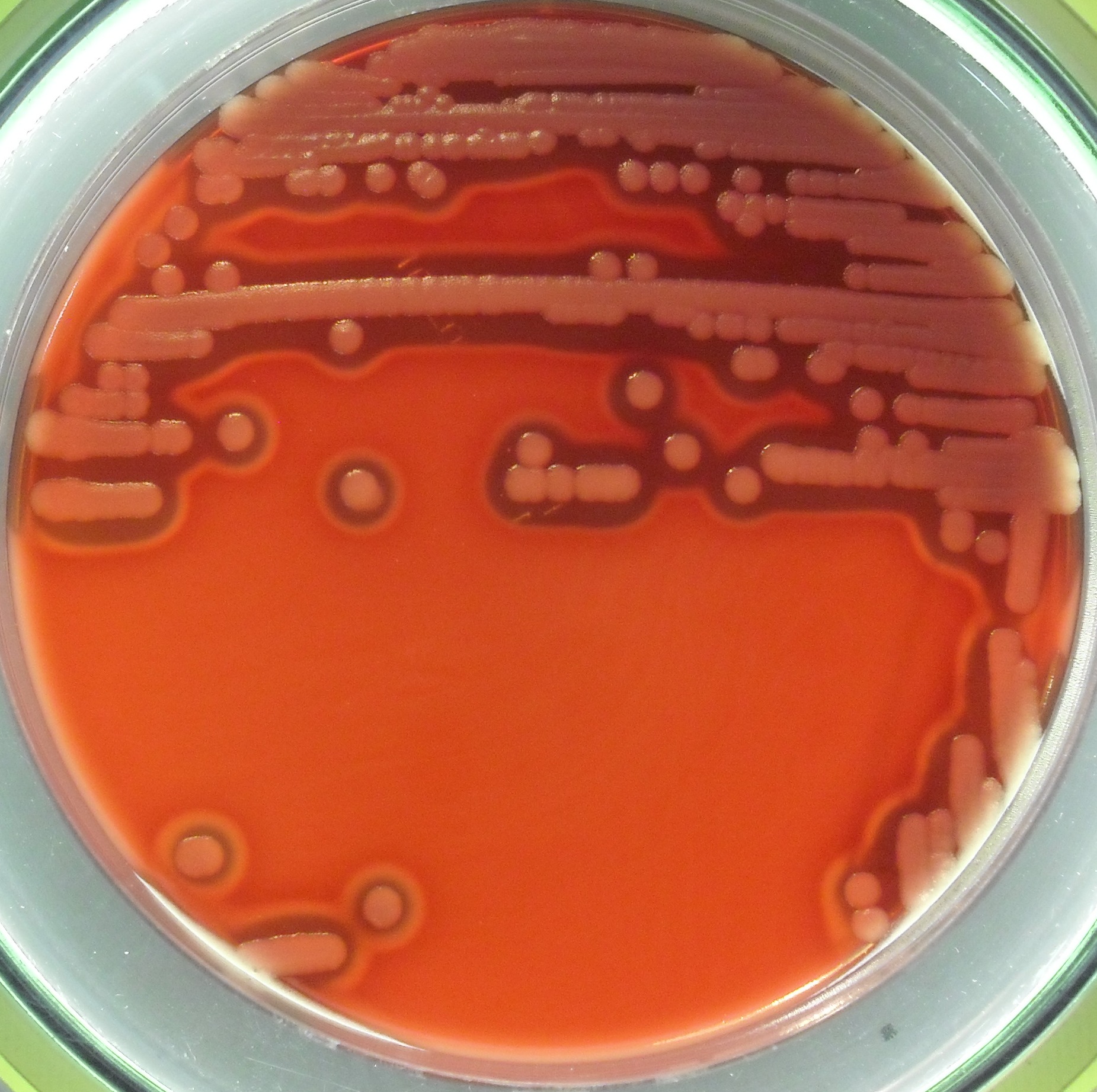
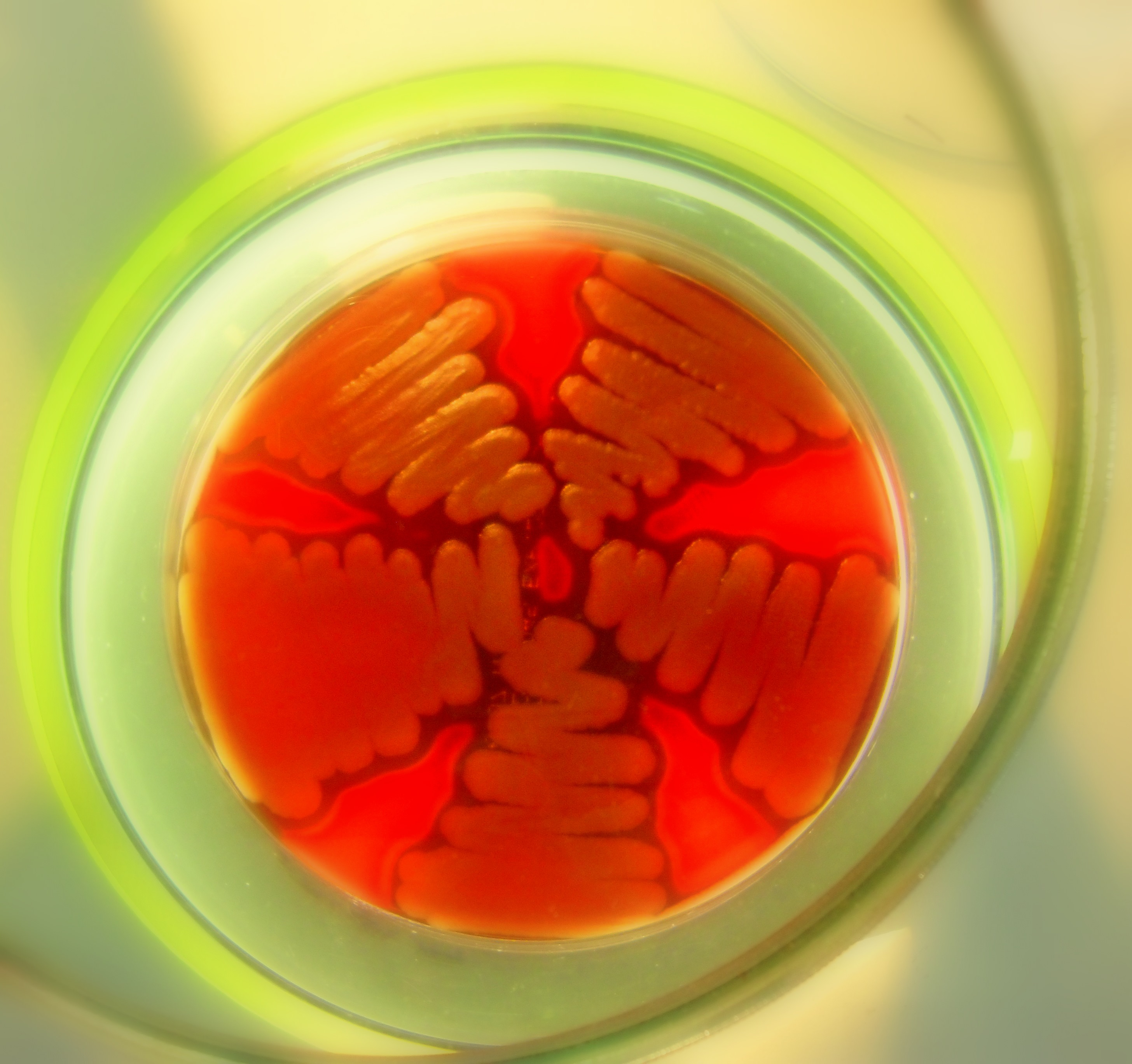
photography series. Experiment to grow slime mold physarum polycephalum (single cell, multi-nuclei organism) inside glass flasks of 100-1000ml, observing the self-organisation and pattern formation of Physarum polycephalum over time. This post was submitted by Theresa Schubert. Fluorescent genetically modified e.coli bacteria. This post was submitted by Ross Colquhoun. A very interesting story about the contribution of microbes to the colours of these ancient artworks.
Thanks to visitor Naomi for the link!
Some initial experiences with photography in a micro laboratory. This post was submitted by Andreia Alves.
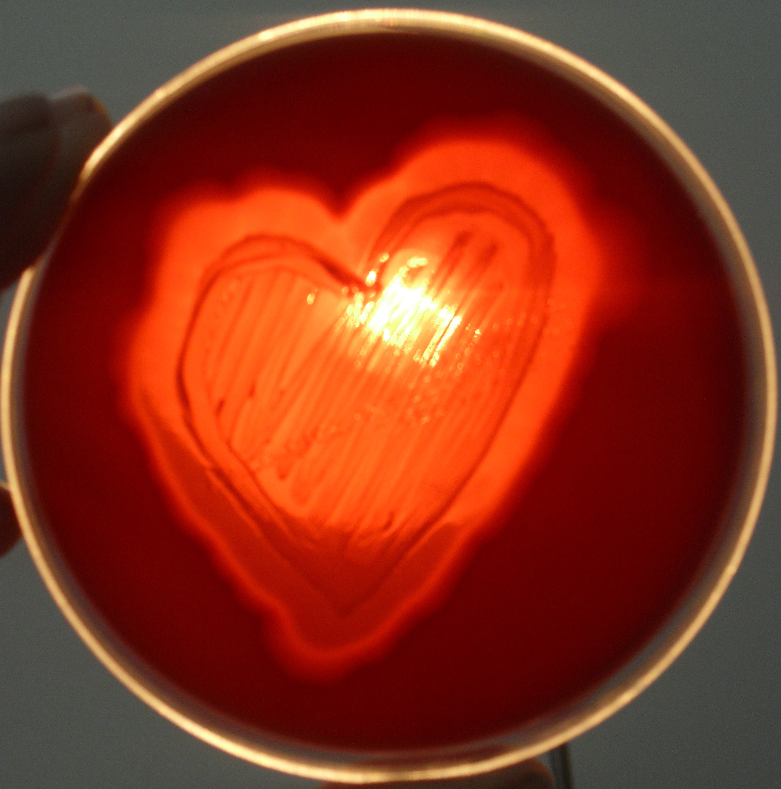
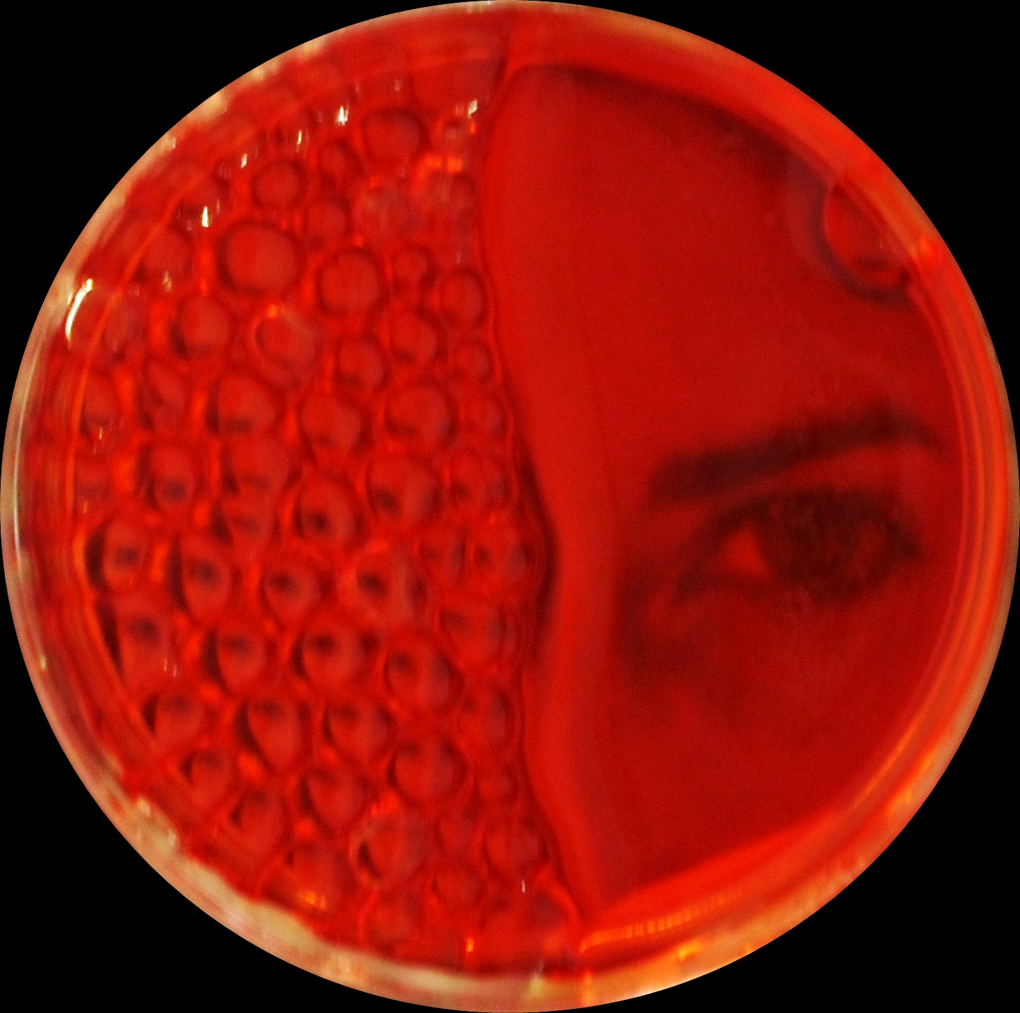
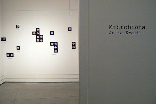
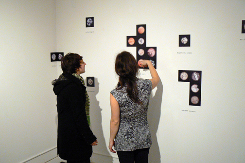
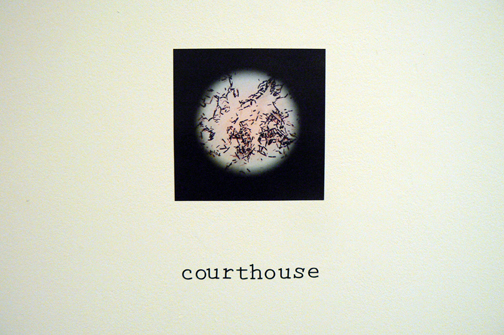
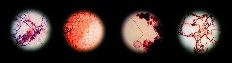
Microbiota | Julia Krolik Microbiota is a photographic installation that maps bacterial life in the city of Kingston. Blending microbiological protocols with artistic vision, the artist reveals invisible yet ubiquitous aspects of our shared environments. This collection of bacterial photographs from various locations in Kingston were sampled, cultured and photographed during the summer of 2010, and aims to raise public awareness of the unseen bacterial environment around us. Julia Krolik received a BScH degree in Biological Sciences from the University of Guelph. A recent placement in Clinical Microbiology combined with Biotechnology studies at St Lawrence College prompted her to raise questions about the way microorganisms are viewed and portrayed in our society. She also co-created Decomposing Pianos, an experimental music project that recently took part in this year’s Tone Deaf festival. This post was submitted by Julia Krolik. Head over to Smithsonian.com and see Painting With Penicillin: Alexander Fleming’s Germ Art by Rob Dunn, which includes a link to Microbialart.com. Two very nice articles with several microbial art images have appeared in the magazines Muy Interesante in Spain (July 2010, p.110-114) and OK in Italy (July 2010, p.104-111). I haven’t been able to find online versions, but check them out if you have access to these publications.
New Scientist has put together a small collection of artworks under a category they call “Eco Art“. This includes an image by Daro Montag, who is a featured artist here on Microbial Art. |
Unique visitors since 10/19/2009 |
|
© 2026 Microbial Art | Powered by WordPress & Atahualpa |
|